Simple Compact Bone Diagram / Compact Bone Tissue Diagram - General Wiring Diagram / In long bones, as you move from the outer cortical compact bone to the inner medullary cavity, the bone transitions to spongy bone.
Simple Compact Bone Diagram / Compact Bone Tissue Diagram - General Wiring Diagram / In long bones, as you move from the outer cortical compact bone to the inner medullary cavity, the bone transitions to spongy bone.. (b) in this micrograph of the osteon, you can clearly see the concentric lamellae and central canals. Compact bone, also called cortical bone, is the hard, stiff, smooth, thin, white bone tissue that surrounds all bones in the human body. In long bones, as you move from the outer cortical compact bone to the inner medullary cavity, the bone transitions to spongy bone. It makes up the outer cortex of all bones and is in immediate contact with the periosteum. Related posts of compact bone diagram labeled anatomy of rib cage.
Compact bone accounts for 80% of the bones in the human body. (b) in this micrograph of the osteon, you can clearly see the concentric lamellae and central canals. Microscopic anatomy bones medical massage physiology pediatric nursing medical knowledge medical anatomy anatomy tutorial structure of bone. In compact bone, these cells are embedded within the solid calcium phosphate matrix of solid bone. Ball and socket joints, like your hip and shoulder joints, are the most mobile type of joint in the human body.
Compact Bones vs. Spongy Bones - Difference and Comparison ... from cdn.shortpixel.ai As shown in figure 2. The inset shows the lamellae of the. It is also called osseous tissue or cortical bone and it provides structure and support for an organism as part of its skeleton, in addition to being a location for the storage of minerals like calcium.about 80% of the weight of the human skeleton comes from. Bone and skeleton fun facts for kids. Anatomy of rib cage 12 photos of the anatomy of rib cage anatomical rib cage necklace, anatomy and physiology of rib cage, anatomy of human rib cage, anatomy of rib cage area, human anatomy rib cage muscles, human anatomy, anatomical rib cage necklace, anatomy and physiology of rib cage, anatomy of human rib cage, anatomy … Those reasons can come off the bones of the diagram. Some, mostly older, compact bone is remodelled to form these haversian systems (or osteons). Bone is made up of two base componets:
Online quiz to learn compact bone diagram;
The remainder is cancellous bone, which has a spongelike appearance with numerous large spaces and is found in the. Anatomy of a long bone anna s anatomy websit. Simple compact bone diagram : Microscopic anatomy bones medical massage physiology pediatric nursing medical knowledge medical anatomy anatomy tutorial structure of bone. Some, mostly older, compact bone is remodelled to form these haversian systems (or osteons). The diagram above shows a longitudinal view of an osteon. These are shown in the figure below. Cancellous or trabecular (spongy) bone; Long bone anatomy structure parts function and fracture types. Compact bone, also called cortical bone, dense bone in which the bony matrix is solidly filled with organic ground substance and inorganic salts, leaving only tiny spaces (lacunae) that contain the osteocytes, or bone cells.compact bone makes up 80 percent of the human skeleton; Compact bone diagram simple diagram system. Some, mostly older, compact bone is remodelled to form these haversian systems (or osteons). (b) in this micrograph of the osteon, you can clearly see the concentric lamellae and central canals.
Bone and skeleton fun facts for kids. Bone is made up of two base componets: Anatomy of a long bone anna s anatomy websit. Cancellous or trabecular (spongy) bone; This type of fibrous joint holds a tooth in place in its socket in the upper and lower jaw.
Cross Section Bone Stock Photos & Cross Section Bone Stock ... from c8.alamy.com Shows compact (cortical) and cancellous (spongy) bone. (b) in this micrograph of the osteon, you can clearly see the concentric lamellae and central canals. Those reasons can come off the bones of the diagram. Terms in this set (8) spongy bone (contains red marrow) compact bone (has osteons) osteon. Compact bone is the denser, stronger of the two types of osseous tissue (figure 6.3.6). Long bone anatomy structure parts function and fracture types. Pig bone diagram wiring diagram, femur bone diagram full human skeleton diagram femur simple compact bone diagram simple diagram system. The main type of bone cell is the osteocyte (bone cell, shown as purple in the diagram).
Long bone diagram compact bone :
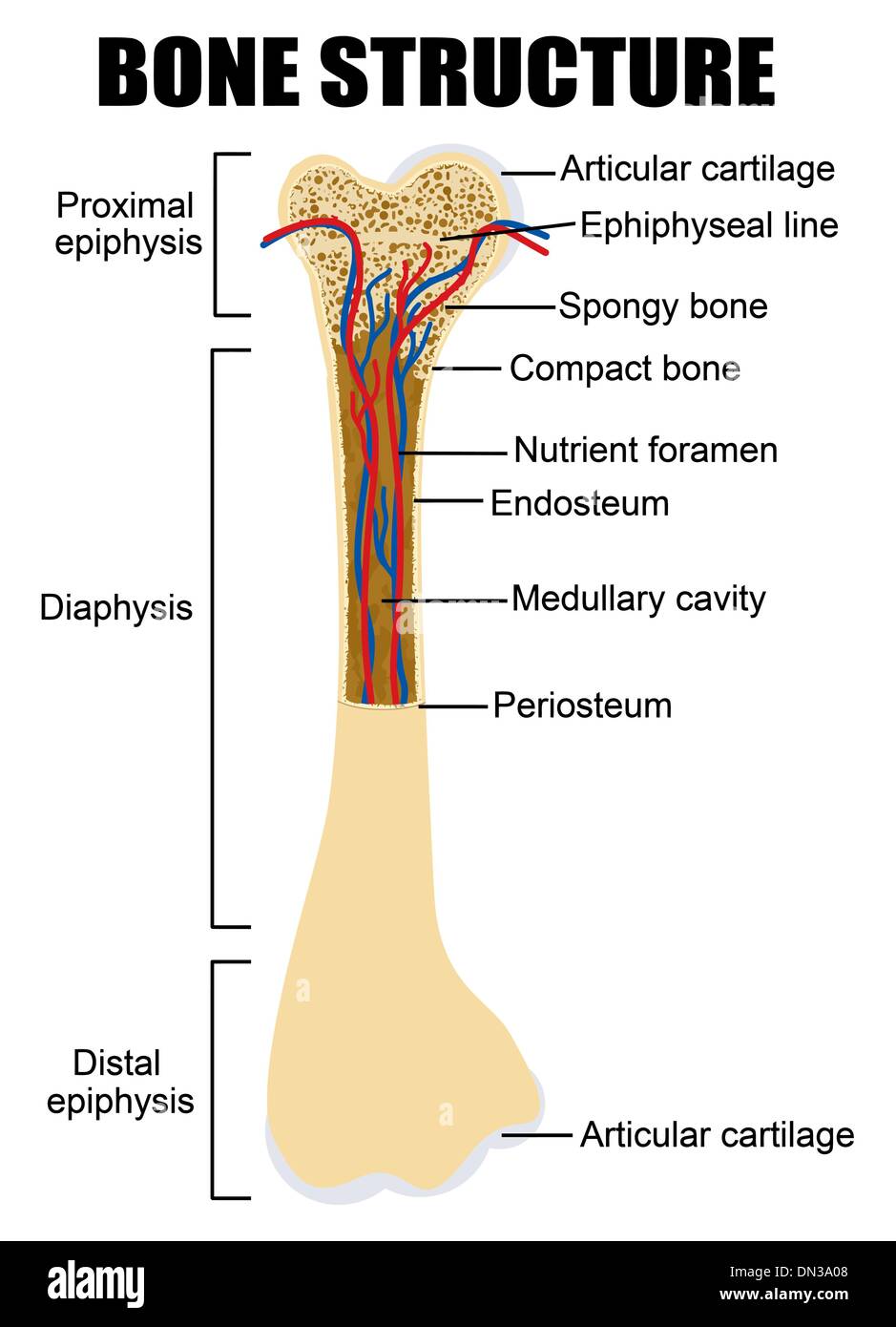
These are shown in the figure below. Compact bone, as opposed to spongy bone, is made of cylindrical units, called osteons, that are tightly formed together. Article by jennifer smith owens. The outer part of a long bone is made of compact bone. Illustration about compact bone, also called cortical bone, is the hard, stiff, smooth, thin, white bone tissue that surrounds all bones in the human body. Cortical bone forms a dense cylinder down the shaft of the bone surrounding the central marrow cavity. Pig bone diagram wiring diagram, femur bone diagram full human skeleton diagram femur simple compact bone diagram simple diagram system. Simple compact bone diagram : The majority of growth during growth spurts is of the long bones. Deep to the compact bone layer is a region of spongy bone where the bone tissue grows in thin columns called. Face bones of the palatine bone diagram. Simple bone diagram wiring diagram. Bone and skeleton fun facts for kids.
The inset shows the lamellae of the. Terms in this set (8) spongy bone (contains red marrow) compact bone (has osteons) osteon. Anatomy of rib cage 12 photos of the anatomy of rib cage anatomical rib cage necklace, anatomy and physiology of rib cage, anatomy of human rib cage, anatomy of rib cage area, human anatomy rib cage muscles, human anatomy, anatomical rib cage necklace, anatomy and physiology of rib cage, anatomy of human rib cage, anatomy … Bone is commonly classified according to its gross appearance as cancellous bone (bone with numerous, macroscopic interconnecting cavities, or trabeculae, also known as spongy or trabecular bone) or compact bone (dense lamellar bone without trabeculae), but both types have the same basic histological structure. Compact bone is made of a matrix of hard mineral salts reinforced with tough collagen fibers.
AccessJ: How to Donate Bone Marrow from 2.bp.blogspot.com Compact bone is the denser, stronger of the two types of osseous tissue (figure 6.3.6). (b) in this micrograph of the osteon, you can clearly see the concentric lamellae and central canals. Many tiny cells called osteocytes live in small spaces in the matrix and help to maintain the strength and integrity of the compact bone. Compact bone is made of a matrix of hard mineral salts reinforced with tough collagen fibers. Long bone anatomy structure parts function and fracture types. However, they do contain osteons, which are like canals, providing passageways through the hard bone matrix. Illustration about compact bone, also called cortical bone, is the hard, stiff, smooth, thin, white bone tissue that surrounds all bones in the human body. Ball and socket joints, like your hip and shoulder joints, are the most mobile type of joint in the human body.
Simple compact bone diagram :
Long bone diagram compact bone : (b) in this micrograph of the osteon, you can clearly see the concentric lamellae and central canals. They are roughly cylindrical, and about 0.2mm wide and a few millimeters long. Compact bone is made of a matrix of hard mineral salts reinforced with tough collagen fibers. Terms in this set (8) spongy bone (contains red marrow) compact bone (has osteons) osteon. Long bones such as the femur contain two distinct morphological types of bone: Cartilage types, their location, bone types, classifications and god knows what else. It makes up the outer cortex of all bones and is in immediate contact with the periosteum. The remainder of the bone is formed by cancellous or spongy bone. Diagram of distinct morphological types of bone. As seen in the image below, compact bone forms the cortex, or hard outer shell of most bones in the body. Compact bone, as opposed to spongy bone, is made of cylindrical units, called osteons, that are tightly formed together. Simple bone diagram wiring diagram.
Osteons are the small units of which the hardest parts of human bones are made compact bone diagram. (b) in this micrograph of the osteon, you can clearly see the concentric lamellae and central canals.


0 Komentar